PhD Research · Optics · Radiation · Microscopy
Exploring how light, X-rays, and electrons interact with matter to reconstruct structure, composition, and function across scales.
Research Areas
Quantitative 3D optical imaging based on holography, angular illumination, and Fourier-space reconstruction of the refractive-index distribution.
Read more →Compact optical-fiber-based radiation detectors for high-resolution dose verification in small and localized radiation therapy fields.
Read more →SEM, EDS, and multi-scale imaging workflows for materials characterization, mineral identification, and porous media analysis.
Read more →Current PhD Research
Recovering the three-dimensional refractive-index distribution of microscopic samples from holographic measurements acquired at multiple illumination angles.
Tomographic Diffractive Microscopy is a quantitative optical imaging technique that reconstructs the internal structure of transparent or weakly scattering samples. Instead of recording only a conventional microscope image, the system measures holograms under multiple illumination angles. Each illumination direction captures a different portion of the sample information in Fourier space.
By combining these measurements, it becomes possible to reconstruct a three-dimensional map of the sample, including variations in refractive index and absorption — without fluorescent labels or sample staining.
Current focus: development of a dual-opposite-view tomographic configuration inspired by 4π microscopy. By combining two opposed transmission views, this approach aims to reduce depth-dependent degradation in the reconstructed volume and partially recover the axial information that is poorly sampled in conventional single-view tomographic microscopy.
Reconstruction pipeline
Hologram acquisition
Extraction of diffracted order
Angular information in reciprocal space
Recovered volume
Radiation Detection
Compact optical sensors for measuring radiation dose in highly localized therapeutic beams.
Modern radiation therapy increasingly uses small and highly focused radiation fields. These fields can deliver dose with strong spatial gradients, which makes accurate dosimetry challenging. Conventional detectors may suffer from volume-averaging effects or may perturb the radiation field they are intended to measure.
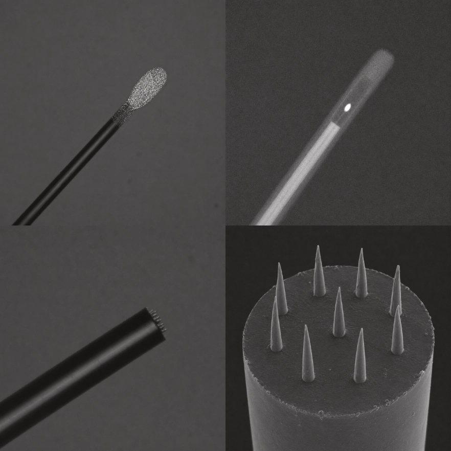
Miniaturized scintillator dosimeters provide an optical solution to this problem. A small scintillating element converts ionizing radiation into light, which is then guided through an optical fiber toward a detector. Because of their compact geometry, these sensors can probe small radiation fields with high spatial resolution.
This research explored the use of a miniaturized inorganic scintillator detector coupled to a narrow optical fiber through a photonic interface. The detector was evaluated under medical photon beams and compared with high-resolution reference probes — including micro-diamond detectors and silicon diodes.
Small sensitive volume
Reduces volume averaging in steep dose gradients.
Optical fiber readout
Light from the scintillator transported through a narrow silica fiber.
Dose verification
Validates delivered dose in small-field radiation therapy.
Reference comparison
Benchmarked against micro-diamond and silicon diode detectors.
Related work: Miniaturized scintillator dosimeter for small field radiation therapy, Physics in Medicine & Biology, 2021.
Scintillator detector · Beam → fiber → photodetector
Measured vs. reference dose profiles
2D dose distribution
Electron Microscopy
Connecting structure, composition, and texture from the millimetre scale down to the micro- and nanoscale using SEM, EDS, and integrated workflows.
Electron microscopy provides a bridge between morphology and composition. Secondary-electron imaging reveals surface topography, backscattered-electron imaging highlights compositional contrast, and energy-dispersive X-ray spectroscopy identifies the elemental distribution of the sample.
This research area focuses on the interpretation of geological and material samples using SEM, EDS, and multi-scale imaging workflows — correlating structural information, mineralogy, porosity, and texture across different spatial scales.
Multi-scale imaging workflows combine X-ray micro-computed tomography, SEM, EDS, QEMSCAN, and image analysis to study complex porous materials such as shales, tight sandstones, and carbonates. The integration of these datasets supports digital rock physics and helps evaluate porosity, pore connectivity, pore-size distribution, and permeability-related features.
In addition to research applications, this line of work includes training activities in scanning electron microscopy. The training focuses on image acquisition, contrast mechanisms, sample preparation, EDS analysis, mineral identification, and interpretation of microstructural features.
SEM · multi-scale imaging workflow
Keywords
My work connects optical physics, numerical reconstruction, radiation detection, and materials characterization.
Publications
Radiation dosimetry · Medical physics
Development and evaluation of a compact inorganic scintillator detector coupled to an optical fiber for high-resolution dose measurements in small photon fields.
View publication →Multi-scale imaging · Digital rock physics
A workflow integrating micro-CT, SEM, QEMSCAN, and image analysis to evaluate mineralogy, pore structure, and petrophysical properties in unconventional reservoir samples.
View publication →PhD research · Optical microscopy
Ongoing development of a dual-opposite-view tomographic diffractive microscopy configuration for improved 3D optical reconstruction beyond the single-view limit.
Learn more →Get in touch
I am open to research collaborations involving quantitative microscopy, optical imaging, electron microscopy, radiation detection, scientific visualization, and computational reconstruction.